An experimental technique for cardiac magnetic resonance (CMR) imaging that is unaffected by cardiac or respiratory motion, and in fact captures such motion as part of the imaging process, could potentially sharpen and simplify CMR procedures, researchers say.
The technique’s inventors, who call it CMR Multitasking, report that it is less technically demanding for CMR technologists compared with current methods and greatly shortens overall scanning time, which is touted as a benefit for both patients and providers.
It doesn’t require workarounds like electrocardiographic (ECG) triggering or patient breath-holds to avoid blurring or other motion artifacts, and it requires only a single dose of contrast agent rather than the multiple doses needed for standard CMR, according to the group.
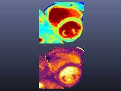
Quantitative T1 (top) and T2 (bottom) maps obtained simultaneously by CMR multitasking, performed without ECG triggering or breath-holds. Courtesy of Anthony G Christodoulou, PhD, and colleagues
“It’s very easy. The technologists just say where they want to image, they press a button, and it goes,” said Anthony G Christodoulou, PhD, Cedars-Sinai Medical Center, Los Angeles, California.
“This should work better for arrhythmia patients, where it’s very hard to trigger based on an ECG, and allows it to work for patients who can’t hold their breath—there are a lot of people who are too sick to do that,” Christodoulou told theheart.org | Medscape Cardiology.
“It’s easier on both patients and the technologist, and allows potentially a push-button acquisition, which is I think the dream for people who worked on cardiac MR for decades. This technique really offers the capability to make that happen,” said Debiao Li, PhD, University of California, Los Angeles, when interviewed.
A report describing the new technique and its use in 10 healthy volunteers and 10 patients with heart disease was published April 9 in Nature Biomedical Engineering, with Christodoulou as lead author and Li as senior author.
MR Multitasking captures conventional two-dimensional images of the heart and simultaneously adds four other “dimensions,” parallel streams of data that represent cardiac motion, the respiratory cycle, and the standard T1-weighted and T2-weighted imagery based on tissue fat and water content.
Data for the added dimensions are all collected during one brief scan and separated out later using software that portrays them as four separate co-registered series of images, for interpretation side by side.
In the reported cases, total imaging time for three separate CMR tests was greatly reduced, achieved sometimes in only 90 seconds, compared to what would have been needed for standard CMR imaging, according to the group.
The technique is under investigation at several centers around the world but isn’t currently used clinically.
Li, Christodoulou, and other coauthors “have a provisional patent application entitled ‘Low-rank tensor imaging for multidimensional cardiovascular MRI’ (USSN 15/495,588).”
Nat Biomed Eng. Published online April 9, 2018. Abstract
Follow Steve Stiles on Twitter: @SteveStiles2. For more from theheart.org | Medscape Cardiology, follow us on Twitter and Facebook.
Tidak ada komentar:
Posting Komentar